The Art and Delivery of Patient Care for Ultrasound
Overview
What is an ultrasound?
Ultrasound (as well called sonography or ultrasonography) is a noninvasive imaging test. An ultrasound moving-picture show is called a sonogram. Ultrasound uses high-frequency sound waves to create existent-fourth dimension pictures or video of internal organs or other soft tissues, such equally claret vessels.
Ultrasound enables healthcare providers to "see" details of soft tissues inside your body without making any incisions (cuts). And unlike X-rays, ultrasound doesn't use radiation.
Although most people associate ultrasound with pregnancy, healthcare providers use ultrasound for many dissimilar situations and to look at several different parts of the inside of your trunk.
How does an ultrasound work?
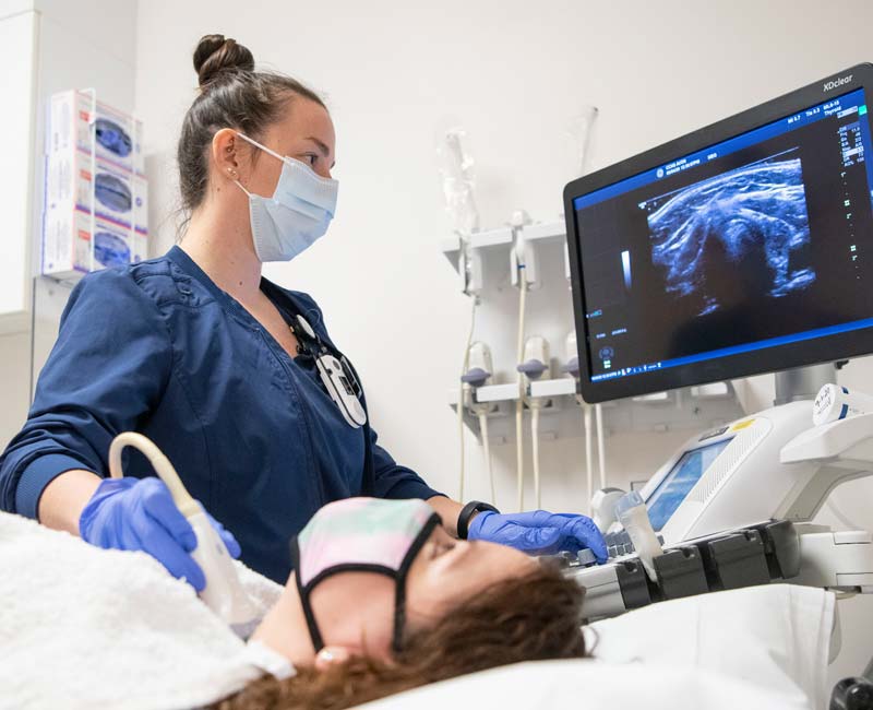
During an ultrasound, a healthcare provider passes a device called a transducer or probe over an area of your body or inside a torso opening. The provider applies a thin layer of gel to your skin so that the ultrasound waves are transmitted from the transducer through the gel and into your body.
The probe converts electric current into loftier-frequency sound waves and sends the waves into your torso's tissue. You tin can't hear the audio waves.
Sound waves bounciness off structures inside your torso and back to the probe, which converts the waves into electrical signals. A calculator and so converts the pattern of electrical signals into real-time images or videos, which are displayed on a computer screen nearby.
What are the dissimilar kinds of ultrasounds?
There are three main categories of ultrasound imaging, including:
- Pregnancy ultrasound (prenatal ultrasound).
- Diagnostic ultrasound.
- Ultrasound guidance for procedures.
Pregnancy ultrasound
Healthcare providers oftentimes utilize ultrasound (often called prenatal or obstetric ultrasound) to monitor you lot and your babe during pregnancy.
Providers use prenatal ultrasound to:
- Confirm that you're pregnant.
- Cheque to see if you lot're pregnant with more than one infant.
- Estimate how long you've been significant and the gestational age of your unborn infant.
- Bank check the fetal growth and position of your unborn baby.
- See the move and center rate of your unborn baby.
- Check for built conditions (birth defects) in your unborn baby's brain, spinal string, heart or other parts of their body.
- Check the amount of amniotic fluid.
Most healthcare providers recommend an ultrasound at 20 weeks pregnant. This test tracks your unborn baby's growth and development during pregnancy. This ultrasound may besides testify your babe'due south biological sex. Tell your technician if you do or exercise not want to know the sex activity.
Your provider may order extra scans to go answers to whatsoever questions or concerns, such equally the potential for congenital conditions.
Diagnostic ultrasound
Providers apply diagnostic ultrasounds to view internal parts of your body to see if something is wrong or not working properly. They tin can help your provider learn more about what's causing a wide range of symptoms, such as unexplained pain, masses (lumps) or what may be causing an aberrant claret test.
For most diagnostic ultrasound exams, the technician places the transducer (probe) on your pare. In some cases, they may demand to place the probe inside your body, such as in your vagina or rectum.
The type of diagnostic ultrasound you accept depends on the details of your example.
Examples of diagnostic ultrasounds include:
- Intestinal ultrasound: An ultrasound probe moves across the skin of your midsection (belly) area. Abdominal ultrasound can diagnose many causes of abdominal pain.
- Kidney (renal) ultrasound: Providers use kidney ultrasound to assess the size, location and shape of your kidneys and related structures, such every bit your ureters and bladder. Ultrasound can detect cysts, tumors, obstructions or infections inside or around your kidneys.
- Breast ultrasound: A breast ultrasound is a noninvasive test to identify breast lumps and cysts. Your provider may recommend an ultrasound later an aberrant mammogram.
- Doppler ultrasound: This is a special ultrasound technique that assesses the move of materials, like claret, in your body. It allows your provider to see and evaluate blood flow through arteries and veins in your body. Doppler ultrasound is ofttimes used every bit part of a diagnostic ultrasound report or as function of a vascular ultrasound.
- Pelvic ultrasound: A pelvic ultrasound looks at the organs in your pelvic area betwixt your lower abdomen (belly) and legs. Some of the pelvic organs include your bladder, prostate, rectum, ovaries, uterus and vagina.
- Transvaginal ultrasound: Your provider inserts a probe into your vaginal canal. It shows reproductive tissues such as your uterus or ovaries. A transvaginal ultrasound is sometimes chosen a pelvic ultrasound because it evaluates structures inside your pelvis (hip bones).
- Thyroid ultrasound: Providers use ultrasound to assess your thyroid, a butterfly-shaped endocrine gland in your neck. Providers tin can measure the size of your thyroid and see if there are nodules or lesions inside the gland.
- Transrectal ultrasound: Your provider inserts an ultrasound probe transducer into your rectum. It evaluates your rectum or other nearby tissues, such as the prostate in people assigned male at nativity.
Ultrasound guidance for procedures
Providers sometimes use ultrasound to perform certain procedures precisely. A common use of ultrasound is to guide needle placement to sample fluid or tissue from:
- Tendons.
- Joints.
- Muscles.
- Cysts or fluid collections.
- Soft-tissue masses.
- Organs (liver, kidney or prostate).
- Transplant organs (liver, kidney or pancreas).
Examples of other procedures that may crave ultrasound guidance include:
- Embryo transfer for in vitro fertilization.
- Nervus blocks.
- Confirming the placement of an IUD (intrauterine device) after insertion.
- Lesion localization procedures.
What is the difference between a 3D ultrasound and a 4D ultrasound?
For ultrasounds during pregnancy, the traditional ultrasound is a two-dimensional (2D) image of your unborn baby. 2D ultrasound produces outlines and apartment-looking images, which allows your healthcare provider to encounter your baby'due south internal organs and structures.
Three-dimensional (3D) ultrasound allows the visualization of some facial features of your baby and perhaps other body parts such as fingers and toes. 4-dimensional (4D) ultrasound is 3D ultrasound in motility. Providers rarely utilize 3D or 4D fetal ultrasound imaging for medical purposes, though it can exist useful in diagnosing a facial or skeletal event. They do, however, utilise 3D ultrasound for other medical purposes, such as evaluating uterine polyps and fibroids.
While ultrasound is by and large considered to be safe with very low risks, the risks may increase with unnecessary prolonged exposure to ultrasound energy or when untrained users operate an ultrasound machine. Because of this, the U.S. Food and Drug Administration (FDA) advises against getting a 3D ultrasound for non-medical reasons such as for "keepsake" moments or entertainment.
Who performs an ultrasound?
A doctor or a healthcare provider called an ultrasound technician or sonographer performs ultrasounds. They're peculiarly trained to operate an ultrasound machine properly and safely.
It's important to always have your ultrasound performed by a medical professional and in a medical facility.
Test Details
How do I ready for an ultrasound?
The preparations will depend on the type of ultrasound you lot're having. Some types of ultrasounds require no preparation at all.
For ultrasounds of the pelvis, including ultrasounds during pregnancy, of the female person reproductive system and of the urinary organisation, you lot may demand to fill up up your bladder by drinking water before the examination.
For ultrasounds of the abdomen, you may need to adjust your diet or fast (not eat or drinkable anything except water) for several hours before your exam.
In any case, your healthcare provider will let you know if y'all need to practice anything special to prepare for your ultrasound. They may give you instructions during an appointment or when scheduling your ultrasound. Instructions may besides exist available in your electronic medical records if you apply such a organization.
What happens during an ultrasound?
Training for an ultrasound varies depending on what body part you'll have scanned. Your provider may inquire you lot to remove certain pieces of clothes or change into a hospital gown.
Ultrasounds that involve applying the transducer (probe) over your pare (not in your body), follow these general steps:
- You'll lie on your side or back on a comfortable tabular array.
- The ultrasound technician volition apply a modest corporeality of h2o-soluble gel on your skin over the area to be examined. This gel doesn't harm your pare or stain your apparel.
- The technician will move a handheld transducer or probe over the gel to get images inside your body.
- The technician may ask you to be very still or to hold your breath for a few seconds to create clearer pictures.
- Once the technician has gotten enough images, they'll wipe off whatever remaining gel on your peel and you'll be done.
An ultrasound examination ordinarily takes 30 minutes to an hour. If y'all take whatsoever questions about your specific type of ultrasound, enquire your healthcare provider.
Is an ultrasound painful?
Ultrasounds that are performed externally (over your skin) are generally not painful. You lot won't feel the audio waves that ultrasound uses. If y'all have to have a full bladder for the process, information technology may be uncomfortable. It may also be uncomfortable to lay on the exam tabular array if you're pregnant.
Ultrasounds that go inside body cavities, such as your vagina or rectum, may exist uncomfortable, just they shouldn't hurt.
Are ultrasounds safe?
Yep, enquiry to engagement has largely shown ultrasound technology to be condom with no harmful side effects. Ultrasound doesn't use radiations, unlike some other medical imaging tests, such equally Ten-rays and CT scans.
Still, all ultrasounds should exist washed by a professional person who has grooming in using this specialized technology safely.
Results and Follow-Upward
When should I know the results of my ultrasound?
The fourth dimension it takes to get your results depends on the type of ultrasound you get. In some cases, such as prenatal ultrasound, your provider may analyze the images and provide results during the test.
In other cases, a radiologist, a healthcare provider trained to supervise and interpret radiology exams, will analyze the images and so ship the study to the provider who requested the examination. Your provider will then share the results with yous or they may be available in your electronic medical tape (if you take an business relationship set up) before your provider reviews the results.
What conditions can be detected by ultrasound?
Ultrasound can help providers diagnose a broad range of medical issues, including:
- Abnormal growths, such as tumors or cancer.
- Claret clots.
- Enlarged spleen.
- Ectopic pregnancy (when a fertilized egg implants exterior of your uterus).
- Gallstones.
- Aortic aneurysm.
- Kidney or float stones.
- Cholecystitis (gallbladder inflammation).
- Varicocele (enlarged veins in the testicles).
What questions should I enquire my healthcare provider well-nigh my ultrasound?
If y'all need an ultrasound, you may want to ask your provider the post-obit questions:
- What blazon of ultrasound do I demand?
- What should I do to prepare for my ultrasound?
- Do I demand any other tests?
- When should I look to get test results?
A note from Cleveland Clinic
Ultrasounds are mutual, rubber and effective imaging tests. Make sure you get an ultrasound from a well-trained professional person (sonographer) who understands how to use this technology properly. If you take any questions nigh your specific ultrasound test, talk to your healthcare provider. They're bachelor to assistance.
sayersuntinxion42.blogspot.com
Source: https://my.clevelandclinic.org/health/diagnostics/4995-ultrasound
0 Response to "The Art and Delivery of Patient Care for Ultrasound"
Post a Comment